AFMシステム
アクセサリ
アプリケーション
お問い合わせ
キャンペーン
「AFMプローブ」 プレゼント
 オックスフォード・インストゥルメンツー事業部ページ

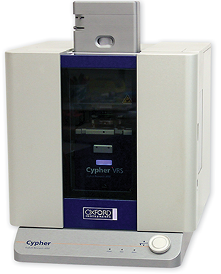
The Cypher VRS AFM is the first and only full-featured video-rate AFM. Finally, researchers can measure nanoscale dynamic processes at video-rate speeds with all of the resolution, versatility, and ease of use that are the hallmarks of an Asylum Research Cypher AFM.
Figure 1B shows just a single frame from this movie. The full movie, however, was collected over a period of about 11 minutes at a rate of 5.8 frames per second. The movie is being played back here at 4X the acquisition rate (i.e. 23.2 fps). The video clearly shows the rows of CTAB hemimicelles on the HOPG surface and the orientation of the domain boundaries. Tapping mode phase data is shown here for optimum contrast.
Figure 2 shows four frames from movie at the indicated times (reference the time stamp on the video, not the player interface). The full movie, however, was collected over a period of about 12 minutes at a rate of 0.48 frames per second. The movie is being played back here at 4X the acquisition rate (i.e. 1.92 fps). Note the two CTAB grains of opposite row orientation (upper-left and lower-right) bounded on the left and right by two grains with parallel orientation. Over time, the two grains narrow at their boundary, eventually separating and drifting apart while the adjacent grains on the left and right merge together. Note the three different row orientations, corresponding to the three directions perpendicular to the HOPG symmetry axes.
Figure 3 shows four frames from this movie. The full movie, however, was collected over a period of about 6 minutes at a rate of 0.95 frames per second. The movie is being played back here at 4X the acquisition rate (i.e. 3.8 fps). The video shows small features that seem to absorb into surrounding grains. The process appears analogous to Ostwald ripening, except in this two-dimensional system instead of a bulk dispersion. In both cases, the feature appears with higher phase contrast and appears less structured than the surrounding hemimicelles.

© オックスフォード・インストゥルメンツ 2026