AFMシステム
アクセサリ
アプリケーション
お問い合わせ
キャンペーン
「AFMプローブ」 プレゼント
 オックスフォード・インストゥルメンツー事業部ページ

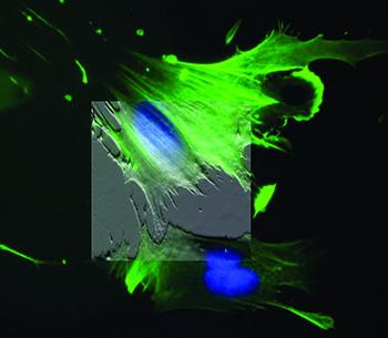
原子間力顕微鏡 (AFM) は細胞生物学研究には不可欠なツールです。 生細胞や固定されていない細胞の情報を3次元形状像で提供することが可能です。しかし、細胞生物学研究における、AFMの最大の特長は、生理学的条件に近い状態(培地中の培養温度37°C)で正確かつ定量的な機械的測定ができることです。フォースマッピングおよびAFMベースのマイクロレオロジー技術を用いて、細胞もしくは基質の弾性・粘弾性応答をルーチンで測定できます。細胞の弾性率は、変性していない細胞、発生・分化・疾患といった異なる状態にある細胞、あるいは薬剤や機械的ストレスなどの刺激に反応する細胞から測定することが可能です。また、基質や細胞の微小環境の弾性率の測定も重要ですが、これは細胞外マトリックス(ECM, extracellular matrix)が、細胞の分化、運命、シグナル伝達、遺伝子転写、癌、心血管疾患およびアポトーシスなどの過程において果たす役割のためです。
倒立型光学顕微鏡(蛍光、共焦点、TIRFなど)と同時に使用した場合、両方の画像診断法から得られるデータを組み合わせて、蛍光標識された構造体をAFM形状像と相互に関連付けすることができます。光学系を使用して、AFMの探針を細胞の特定領域をプローブするように移動させることが可能です。これは特に、イメージングが困難な細胞を対象とする場合は、極めて重要になり得ます。最後に、AFMは細胞へ機械的刺激を与え、関連する応答(例えば、イオン処理、膜電位変化など)を光学的に記録し、生細胞および組織における機械的シグナル伝達を理解することが可能です。
画像をクリックするとダウンロードできます。
"Peptide density targets and impedes triple negative breast cancer metastasis," D. Liu, P. Guo, C. McCarthy, B. Wang, Y. Tao, and D. Auguste, Nat. Commun. 9, 2612 (2018). https://doi.org/10.1038/s41467-018-05035-5
"Direct micropatterning of extracellular matrix proteins on functionalized polyacrylamide hydrogels shows geometric regulation of cell–cell junctions," B. Sarker, C. Walter, and A. Pathak, ACS Biomater. Sci. Eng. 4, 2340 (2018). https://doi.org/10.1021/acsbiomaterials.8b00331
"Measuring nanoscale viscoelastic parameters of cells directly from AFM force-displacement curves," Y. M. Efremov, W.-H. Wang, S. D. Hardy, R. L. Geahlen, and A. Raman, Sci. Rep. 7, 1541 (2017). https://doi.org/10.1038/s41598-017-01784-3
"3D cell bioprinting of self-assembling peptide-based hydrogels," B. Raphael, T. Khalil, V. L. Workman, A. Smith, C. P. Brown, C. Streuli, A. Saiani, and M. Domingos, Mater. Lett. 190, 103 (2017). https://doi.org/10.1016/j.matlet.2016.12.127
"T cell activation requires force generation," K. H. Hu and M. J. Butte, J. Cell Biol. 213, 535 (2016). https://doi.org/10.1083/jcb.201511053
"Mechanical heterogeneities in the subendothelial matrix develop with age and decrease with exercise," J. C. Kohn, A. Chen, S. Cheng, D. R. Kowal, M. R. King, and C. A. Reinhart-King, J. Biomech. 49, 1447 (2016). https://doi.org/10.1016/j.jbiomech.2016.03.016
"Human breast cancer invasion and aggression correlates with ECM stiffening and immune cell infiltration," I. Acerbi, L. Cassereau, I. Dean, Q. Shi, A. Au, C. Park, Y. Y. Chen, J. Liphardt, E. S. Hwang, and V. M. Weaver, Integr. Biol. 7, 1120 (2015). https://doi.org/10.1039/c5ib00040h
"Nanomechanics of cells and biomaterials studied by atomic force microscopy," J. I. Kilpatrick, I. Revenko, and B. J. Rodriguez, Adv. Healthcare Mater. 4, 2456 (2015). https://doi.org/10.1002/adhm.201500229
"Tissue mechanics modulate microRNA-dependent PTEN expression to regulate malignant progression," J. K. Mouw, Y. Yui, L. Damiano, R. O. Bainer, J. N. Lakins, I. Acerbi, G. Ou, A. C. Wijekoon, K. R. Levental, P. M. Gilbert, E. S. Hwang, Y.-Y. Chen, and V. M. Weaver, Nat. Med. 20, 360 (2014). https://doi.org/10.1038/nm.3497
"Atomic force microscopy-based microrheology reveals significant differences in the viscoelastic response between malign and benign cell lines," J. Rother, H. Nöding, I. Mey, and A. Janshoff, Open Biol. 4, 140046 (2014). https://doi.org/10.1098/rsob.140046
"A physical sciences network characterization of non-tumorigenic and metastatic cells," The Physical Sciences – Oncology Centers Network (D. B. Agus et al.), Sci. Rep. 3, 1449 (2013). https://doi.org/10.1038/srep01449
"Comparison of the viscoelastic properties of cells from different kidney cancer phenotypes measured with atomic force microscopy," L. M. Rebelo, J. S. de Sousa, J. Mendes Filho, and M. Radmacher, Nanotechnology 24, 055102 (2013). https://doi.org/10.1088/0957-4484/24/5/055102
"Elasticity maps of living neurons measured by combined fluorescence and atomic force microscopy," E. Spedden, J. D. White, E. N. Naumova, D. L. Kaplan, and C. Staii, Biophys. J. 103, 868 (2012). https://doi.org/10.1016/j.bpj.2012.08.005
"Cell stiffness is a biomarker of the metastatic potential of ovarian cancer cells," W. Xu, R. Mezencev, B. Kim, L. Wang, J. McDonald, and T. Sulchek, PLoS One 7, e46609 (2012). https://doi.org/10.1371/journal.pone.0046609
"In situ force mapping of mammary gland transformation," J. I. Lopez, I. Kang, W.-K. You, D. M. McDonald, and V. M. Weaver, Integr. Biol. 3, 910 (2011). https://doi.org/10.1039/c1ib00043h
"Mapping nanomechanical properties of live cells using multi-harmonic atomic force microscopy," A. Raman, S. Trigueros, A. Cartagena, A. P. Z. Stevenson, M. Susilo, E. Nauman, and S. A. Contera, Nat. Nanotechnol. 6, 809 (2011). https://doi.org/10.1038/nnano.2011.186
"Detection of single-molecule H2O2 signalling from epidermal growth factor receptor using fluorescent single-walled carbon nanotubes," H. Jin, D. A. Heller, M. Kalbacova, J.-H. Kim, J. Zhang, A. A. Boghossian, N. Maheshri, and M. S. Strano, Nat. Nanotechnol. 5, 302 (2010). https://doi.org/10.1038/nnano.2010.24
"Nanotopography-induced changes in focal adhesions, cytoskeletal organization, and mechanical properties of human mesenchymal stem cells," E. K. Yim, E. M. Darling, K. Kulangara, F. Guilak, and K. W. Leong, Biomaterials 31, 1299 (2010). https://doi.org10.1016/j.biomaterials.2009.10.037
"Impact of actin rearrangement and degranulation on the membrane structure of primary mast cells: A combined atomic force and laser scanning confocal microscopy investigation," Z. Deng, T. Zink, H. Chen, D. Walters, F. Liu, and G. Liu, Biophys. J. 96, 1629 (2009). https://doi.org/10.1016/j.bpj.2008.11.015
"Control of myocyte remodeling in vitro with engineered substrates," N. A. Geisse, S. P. Sheehy, and K. K. Parker, In Vitro Cell. Dev. Biol.–Animal 45, 343 (2009). https://doi.org/10.1007/s11626-009-9182-9
"Intermediate filament-like proteins in bacteria and a cytoskeletal function in Streptomyces," S. Bagchi, H. Tomenius, L. M. Belova, and N. Ausmees, Mol. Microbiol. 70, 1037 (2008). https://doi.org/10.1111/j.1365-2958.2008.06473.x
"A cell nanoinjector based on carbon nanotubes," X. Chen, A. Kis, A. Zettl, and C. R. Bertozzi, Proc. Natl. Acad. Sci. U. S. A. 104, 8218 (2007). https://doi.org/10.1073/pnas.0700567104
"Matrix elasticity directs stem cell lineage specification," A. J. Engler, S. Sen, H. L. Sweeney, and D. E. Discher, Cell 126, 677 (2006). https://doi.org/10.1016/j.cell.2006.06.044
"Indentation and adhesive probing of a cell membrane with AFM: Theoretical model and experiments," S. Sen, S. Subramanian, and D. E. Discher, Biophys. J. 89, 3203 (2005). https://doi.org/10.1529/biophysj.105.063826
"Myotubes differentiate optimally on substrates with tissue-like stiffness: pathological implications for soft or stiff microenvironments," A. J. Engler, M. A. Griffin, S. Sen, C. G. Bönnemann, H. L. Sweeney, and D. E. Discher, J. Cell Biol. 166, 877 (2004). https://doi.org/10.1083/jcb.200405004

© オックスフォード・インストゥルメンツ 2026