AFMシステム
アクセサリ
アプリケーション
お問い合わせ
キャンペーン
「AFMプローブ」 プレゼント
 オックスフォード・インストゥルメンツー事業部ページ

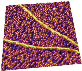
原子間力顕微鏡 (AFM) は、生理学的条件に近い状態で分子の構造を解明することができる強力なツールです。 サンプルを自然な状態、つまり生体関連温度において常に水を含んだ状態でイメージングすることが可能です。サンプルに対して、固定・コーティング・脱水といった追加処理は必要ありません。AFMの主な特長は、サンプルの動的変化をモニターできることです。サンプル調整が最小限で済むため、分子間の相互作用および外部因子に対する分子の応答を観察することが可能です。AFMのもう一つの機能は、分子の機械特性の測定が可能なことです。 ピコニュートンの力を検出できるため、分子内および分子間力の測定が可能となります。これにより、タンパク質の構造や、その構造をほどくのに必要な力など、タンパク質の動態に関する理解を深めることができます。
AFMに関する技術的なお問い合わせ画像をクリックするとダウンロードできます。
"Multifrequency AFM reveals lipid membrane mechanical properties and the effect of cholesterol in modulating viscoelasticity," Z. Al-Rekabi and S. Contera, Proc. Natl. Acad. Sci. U.S.A. 115, 2658 (2018). https://doi.org/10.1073/pnas.1719065115
"High-resolution AFM structure of DNA G-wires in aqueous solution," K. Bose, C. J. Lech, B. Heddi, and A. T. Phan, Nat. Commun. 9, 1959 (2018). https://doi.org/10.1038/s41467-018-04016-y
"Controlling the mechanoelasticity of model biomembranes with room-temperature ionic liquids," C. Rotella, P. Kumari, B. J. Rodriguez, S. P. Jarvis, and A. Benedetto, Biophys. Rev. 10, 751 (2018). https://doi.org/10.1007/s12551-018-0424-5
"A novel pathway for amyloids self-assembly in aggregates at nanomolar concentration mediated by the interaction with surfaces," S. Banerjee, M. Hashemi, Z. Lv, S. Maity, J. C. Rochet, and Y. L. Lyubchenko, Sci. Rep. 7, 45592 (2017). https://doi/org/10.1038/srep45592
"Endothelial glycocalyx-mediated nitric oxide production in response to selective AFM pulling," A. M. W. Bartosch, R. Mathews, and J. M. Tarbell, Biophys. J. 113, 101 (2017). https://doi.org/10.1016/j.bpj.2017.05.033
"DNA nanostructures-mediated molecular imprinting lithography," C. Tian, H. Kim, W. Sun, Y. Kim, P. Yin, and H. Liu, ACS Nano 11, 227 (2017). https://doi.org/10.1021/acsnano.6b04777
"Self-organized architectures from assorted DNA-framed nanoparticles," W. Liu, J. Halverson, Y. Tian, A. V. Tkachenko, and O. Gang, Nat. Chem. 8, 867 (2016). https://doi.org/10.1038/nchem.2540
"TRF2-mediated control of telomere DNA topology as a mechanism for chromosome-end protection," D. Benarroch-Popivker, S. Pisano, A. Mendez-Bermudez, L. Lototska, P. Kaur, S. Bauwens, N. Djerbi, C. M. Latrick, V. Fraisier, B. Pei, A. Gay, E. Jaune, K. Foucher, J. Cherfils-Vicini, E. Aeby, S. Miron, A. Londoño-Vallejo, J. Ye, M.-H. Le Du, H. Wang, E. Gilson, and M.-J. Giraud-Panis, Mol. Cell 61, 274 (2016). http://dx.doi.org/10.1016/j.molcel.2015.12.009
"Visualizing the path of DNA through proteins using DREEM imaging," D. Wu, P. Kaur, Z. M. Li, K. C. Bradford, H. Wang, and D. A. Erie, Mol. Cell 61, 315 (2016). https://doi.org/10.1016/j.molcel.2015.12.012
"Titin domains progressively unfolded by force are homogenously distributed along the molecule," P. Bianco, Z. Mártonfalvi, K. Naftz, D. Koszegi, and M. Kellermayer, Biophys. J. 109, 340 (2015). https://doi.org/10.1016/j.bpj.2015.06.002
"Direct observation of the reversible two‐state unfolding and refolding of an α/β protein by single‐molecule atomic force microscopy," C. He, C. Hu, X. Hu, X. Hu, A. Xiao, T. T. Perkins, and H. Li, Angew. Chem. Intl. Ed. 54, 9921 (2015). https://doi.org/10.1002/anie.201502938
"Effect of the interaction of the amyloid β (1–42) peptide with short single-stranded synthetic nucleotide sequences: Morphological characterization of the inhibition of fibrils formation and fibrils disassembly," J. N. Abraham, D. Kedracki, E. Prado, C. Gourmel, P. Maroni, and C. Nardin, Biomacromolecules 15, 3253 (2014). https://doi.org/10.1021/bm501004q
"Multiparametric high-resolution imaging of native proteins by force-distance curve–based AFM," M. Pfreundschuh, D. Martinez-Martin, E. Mulvihill, S. Wegmann, and D. J. Muller, Nat. Protoc. 9, 1113 (2014). https://doi.org/10.1038/nprot.2014.070
"The nanomechanical properties of lipid membranes are significantly influenced by the presence of ethanol," F. W. S. Stetter and T. Hugel, Biophys. J. 104, 1049 (2013). https://doi.org/10.1016/j.bpj.2013.01.021
"Non-DLVO adhesion of F-specific RNA bacteriophages to abiotic surfaces: Importance of surface roughness, hydrophobic and electrostatic interactions," C. Dika, M. Ly-Chatain, G. Francius, J. Duval, and C. Gantzer, Colloids Surf. A 435, 178 (2013). https://doi.org/10.1016/j.colsurfa.2013.02.045
"Distinct annular oligomers captured along the assembly and disassembly pathways of transthyretin amyloid protofibrils," R. H. Pires, Á. Karsai, M. J. Saraiva, A. M. Damas, and M. S. Z. Kellermayer, PLoS One 7, e44992 (2012). https://doi.org/10.1371/journal.pone.0044992
"Surface characterization and AFM imaging of mixed fibrinogen–surfactant films," N. Hassan, J. Maldonado-Valderrama, A. P. Gunning, V. J. Morris, and J. M. Ruso, J. Phys. Chem. B 115, 6304 (2011). https://doi.org/10.1021/jp200835j
"Site-specific attachment of proteins onto a 3D DNA tetrahedron through backbone-modified phosphorothioate DNA," N. Y. Wong, C. Zhang, L. H. Tan, and Y. Lu, Small 7, 1427 (2011). https://doi.org/10.1002/smll.201100140
"Tuning the elastic modulus of hydrated collagen fibrils," C. A. Grant, D. J. Brockwell, S. E. Radford, and N. H. Thomson, Biophys. J. 97, 2985 (2009). https://doi.org/10.1016/j.bpj.2009.09.010
"Stepwise dynamics of epitaxially growing single amyloid fibrils," M. S. Z. Kellermayer, A. Karsai, M. Benke, K. Soos, and B. Penke, Proc. Natl. Acad. Sci. U.S.A. 105, 141 (2007). https://doi.org/10.1073/pnas.0704305105
"Packing density and structural heterogeneity of insulin amyloid fibrils measured by AFM nanoindentation," S. Guo, and B. B. Akhremitchev, Biomacromolecules 7, 1630 (2006). https://doi.org/10.1021/bm0600724
"Temperature softening of a protein in single-molecule experiments," M. Schlierf and M. Rief, J. Mol. Biol. 354, 497 (2005). https://doi.org/10.1016/j.jmb.2005.09.070
"Reversible mechanical unzipping of amyloid β-fibrils," M. S. Z. Kellermayer, L. Grama, Á. Karsai, A. Nagy, A. Kahn, Z. L. Datki, and B. Penke, J. Biol. Chem. 280, 8464 (2004). https://doi.org/10.1074/jbc.m411556200
"Segmented nanofibers of spider dragline silk: Atomic force microscopy and single-molecule force spectroscopy," E. Oroudjev, J. Soares, S. Arcidiacono, J. B. Thompson, S. A. Fossey, and H. G. Hansma, Proc. Natl. Acad. Sci. U.S.A. 99, 6460 (2002). https://doi.org/10.1073/pnas.082526499
"Can non-mechanical proteins withstand force? Stretching barnase by atomic force microscopy and molecular dynamics simulation," R. B. Best, B. Li, A. Steward, V. Daggett, and J. Clarke, Biophys. J. 81, 2344 (2001). https://doi.org/10.1016/s0006-3495(01)75881-x

© オックスフォード・インストゥルメンツ 2026