AFMシステム
アクセサリ
アプリケーション
お問い合わせ
キャンペーン
「AFMプローブ」 プレゼント
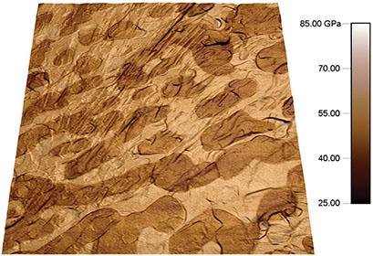
ナノスケールでの機械特性は、多くのアプリケーションにおいて非常に重要です。原子間力顕微鏡(AFM, atomic force microscopy)は、これらの特性を計測できる限られたツール中の1つです。 アサイラム・リサーチAFM用のNanomechPro™ ツールキットを使用することで、細胞からセラミックまで、あらゆる機械特性をナノスケールで測定することが可能になります。このツールキットに含まれるテクニックにより、弾性・粘性、接着力、硬度など、広範囲にわたるナノメカニカル挙動を正確に評価することができます。NanomechProツールキット内の複数のテクニックは、異なるアプリケーションに対して高い柔軟性を提供し、得られた結果を比較することで、より深い知見を得ることができます。NanomechProツールキットには、Cypher™ ファミリー・MFP-3D™ ファミリーのいずれのAFMにも対応した機能が含まれており、多数の特性を高速で測定できる独自のモードと一緒に使用できます。
Cypher AFM用のレーザー干渉変位計測オプション(IDS, Interferometric Displacement Sensor)を使用すると、ナノメカニカル特性評価モードにおいて、より定量的かつ詳細な計測が可能です。従来の光てこによるたわみ検出法(OBD, optical beam deflection)では、カンチレバー形状が予測された形やモデル型からそれた場合、OBD信号が誤って解釈される可能性があります。 これに対して、IDSはカンチレバーの振幅・たわみの絶対値を提供するため、マルチ周波数テクニック、モード形のマッピング、チップ - サンプル接触機構、およびオンオフ共振接触テクニックの精度を向上させます。下の技術資料のタブをクリックしてIDSオプションのデータシートをダウンロードしていただけます。
AFMに関する技術的なお問い合わせ画像をクリックするとダウンロードできます。
"Probing the swelling-dependent mechanical and transport properties of polyacrylamide hydrogels through AFM-based dynamic nanoindentation," Y. Lai and Y. Hu, Soft Matter 14, 2619 (2018). https://doi.org/10.1039/c7sm02351k
"Controlling the mechanoelasticity of model biomembranes with room-temperature ionic liquids," C> Rotella, P. Kumari, B. J. Rodriguez, S. P. Jarvis, and A. Benedetto, Biophys. Rev. 10, 751 (2018). https://doi.org/10.1007/s12551-018-0424-5
"原子間力顕微鏡を利用したナノ力学物性測定技術," 谷口 幸範, 日本ゴム協会誌, 2017 年 90 巻 12 号 p. 577-582. https://doi.org/10.2324/gomu.90.577
"Tendon exhibits complex poroelastic behavior at the nanoscale as revealed by high-frequency AFM-based rheology," B. K. Connizzo and A. J. Grodzinsky, J. Biomech. 54, 11 (2017). https://doi.org/10.1016/j.jbiomech.2017.01.029
"Polymer nanomechanics: Separating the size effect from the substrate effect in nanoindentation," L. Li, L. M. Encarnacao, and K. A. Brown, Appl. Phys. Lett. 110, 043105 (2017). https://doi.org/10.1063/1.4975057
"Mechanical properties of highly porous super liquid‐repellent surfaces," M. Paven, R. Fuchs, T. Yakabe, D.,Vollmer, M. Kappl, A. N. Itakura, and H.-J. Butt, Adv. Funct. Mater. 26, 4914 (2016). https://doi.org/10.1002/adfm.201600627
"Practical loss tangent imaging with amplitude-modulated atomic force microscopy," R. Proksch, M. Kocun, D. Hurley, M. Viani, A. Labuda, W. Meinhold, and J. Bemis, J. Appl. Phys. 119, 134901 (2016). https://doi.org/10.1063/1.4944879
"Fast, quantitative AFM nanomechanical measurements using AM-FM Viscoelastic Mapping mode," D. Hurley, M. Kocun, I. Revenko, B. Ohler, and R. Proksch, Microscopy and Analysis 29, 9 (2015). Download Here
"Contact resonance atomic force microscopy imaging in air and water using photothermal excitation," M. Kocun, A. Labuda, A. Gannepalli, and R. Proksch, Rev. Sci. Instrum. 86, 083706 (2015). https://doi.org/10.1063/1.4928105
"Predictive modelling-based design and experiments for synthesis and spinning of bioinspired silk fibres," S. Lin, S. Ryu, O. Tokareva, G. Gronau, M. M. Jacobsen, W. Huang, D. J. Rizzo, D. Li, C. Staii, N. M. Pugno, J. Y. Wong, D. L. Kaplan, and M. J. Buehler, Nat. Comm. 6, 6892 (2015). http://doi.org/10.1038/ncomms7892
"Nano-rheology of hydrogels using direct drive force modulation atomic force microscopy," P. C. Nalam, N. N. Gosvami, M. A. Caporizzo, R. J. Composto, and R. W. Carpick, Soft Matter 11, 8165 (2015). https://doi.org/10.1039/c5sm01143d
"Fast nanomechanical spectroscopy of soft matter," E. T. Herruzo, A. P. Perrino, and R. Garcia, Nat. Commun. 5, 3126 (2014). https://doi.org/10.1038/ncomms4126
"High intrinsic mechanical flexibility of mouse prion nanofibrils revealed by measurements of axial and radial Young's moduli," G. Lamour, C. K. Yip, H. Li, and J. Gsponer, ACS Nano 8, 3851 (2014). https://doi.org/10.1021/nn5007013
"Quantifying cell-to-cell variation in power-law rheology," P. Cai, Y. Mizutani, M. Tsuchiya, J. M. Maloney, B. Fabry, K. J. V. Vliet, and T. Okajima, Biophys. J. 105, 1093 (2013). https://doi.org/10.1016/j.bpj.2013.07.035
"Nanomechanical mapping of soft matter by bimodal force microscopy," R. Garcia and R. Proksch, Eur. Polym. J. 49, 1897 (2013). https://doi.org/10.1016/j.eurpolymj.2013.03.037
"Loss tangent imaging: Theory and simulations of repulsive-mode tapping atomic force microscopy," R. Proksch and D. G. Yablon, Appl. Phys. Lett. 100, 073106 (2012). https://doi.org/10.1063/1.3675836
"Mapping nanoscale elasticity and dissipation using dual frequency contact resonance AFM," A. Gannepalli, D. G. Yablon, A. H. Tsou, and R. Proksch, Nanotechnology 22, 355705 (2011). https://doi.org/10.1088/0957-4484/22/35/355705
"Mapping nanomechanical properties of live cells using multi-harmonic atomic force microscopy," A. Raman, S. Trigueros, A. Cartagena, A. P. Z. Stevenson, M. Susilo, E. Nauman, and S. A. Contera, Nat. Nanotechnol. 6, 809 (2011). https://doi.org/10.1038/nnano.2011.186
"Viscoelastic property mapping with contact resonance force microscopy," J. P. Killgore, D. G. Yablon, A. Tsou, A. Gannepalli, P. Yuya, J. Turner, R. Proksch, and D. C. Hurley, Langmuir 27, 13983 (2011). https://doi.org/10.1021/la203434w
"Mechanical properties of face-centered cubic supercrystals of nanocrystals," E. Tam, P. Podsiadlo, E. Shevchenko, D. F. Ogletree, M.-P. Delplancke-Ogletree, and P. D. Ashby, Nano Lett. 10, 2363 (2010). https://doi.org/10.1021/nl1001313
"Tuning the elastic modulus of hydrated collagen fibrils," C. A. Grant, D. J. Brockwell, S. E. Radford, and N. H. Thomson, Biophys. J. 97, 2985 (2009). https://doi.org/10.1016/j.bpj.2009.09.010
"Vascular smooth muscle cell durotaxis depends on substrate stiffness gradient strength," B. C. Isenberg, P. A. DiMilla, M. Walker, S. Kim, and J. Y. Wong, Biophys. J. 97, 1313 (2009). https://doi.org/10.1016/j.bpj.2009.06.021
"Surface viscoelasticity of individual gram-negative bacterial cells measured using atomic force microscopy," V. Vadillo-Rodriguez, T. J. Beveridge, and J. R. Dutcher, J. Bacteriol. 190, 4225-4232 (2008). https://doi.org/10.1128/jb.00132-08
"A thin-layer model for viscoelastic, stress-relaxation testing of cells using atomic force microscopy: Do cell properties reflect metastatic potential?" E. M. Darling, S. Zauscher, J. A. Block, and F. Guilak, Biophys. J. 92, 1784 (2007). https://doi.org/10.1529/biophysj.106.083097
"Packing density and structural heterogeneity of insulin amyloid fibrils measured by AFM nanoindentation," S. Guo, and B. B. Akhremitchev, Biomacromolecules 7, 1630 (2006). https://doi.org/10.1021/bm0600724
"Multifrequency, repulsive-mode amplitude-modulated atomic force microscopy," R. Proksch, Appl. Phys. Lett. 89, 113121 (2006). https://doi.org/10.1063/1.2345593
© オックスフォード・インストゥルメンツ 2026